Lung Bone Anatomy
Download this free Lung Bone Anatomy and use it right away. Optimized for A4 and Letter paper, all 35 designs are ready to print without editing software. No sign-up required.
How to Use This Lung Bone Anatomy
- Browse the collectionScroll through the Lung Bone Anatomy designs above and click any image to open it full size.
- Download the imageHit the Download button to save the full-resolution file to your device.
- Print on standard paperUse A4 or Letter paper. Select 'Fit to page' in your printer settings to ensure nothing is cut off.
- Use immediatelyNo editing, software, or account needed — it's ready the moment it comes out of the printer.
More Lung Bone Anatomy Templates
 Desmoplastic Fibroma In The Proximal Femur A Case Report With Long
 Radiographs Demonstrating An Expansile Lytic Lesion Involving The Whole
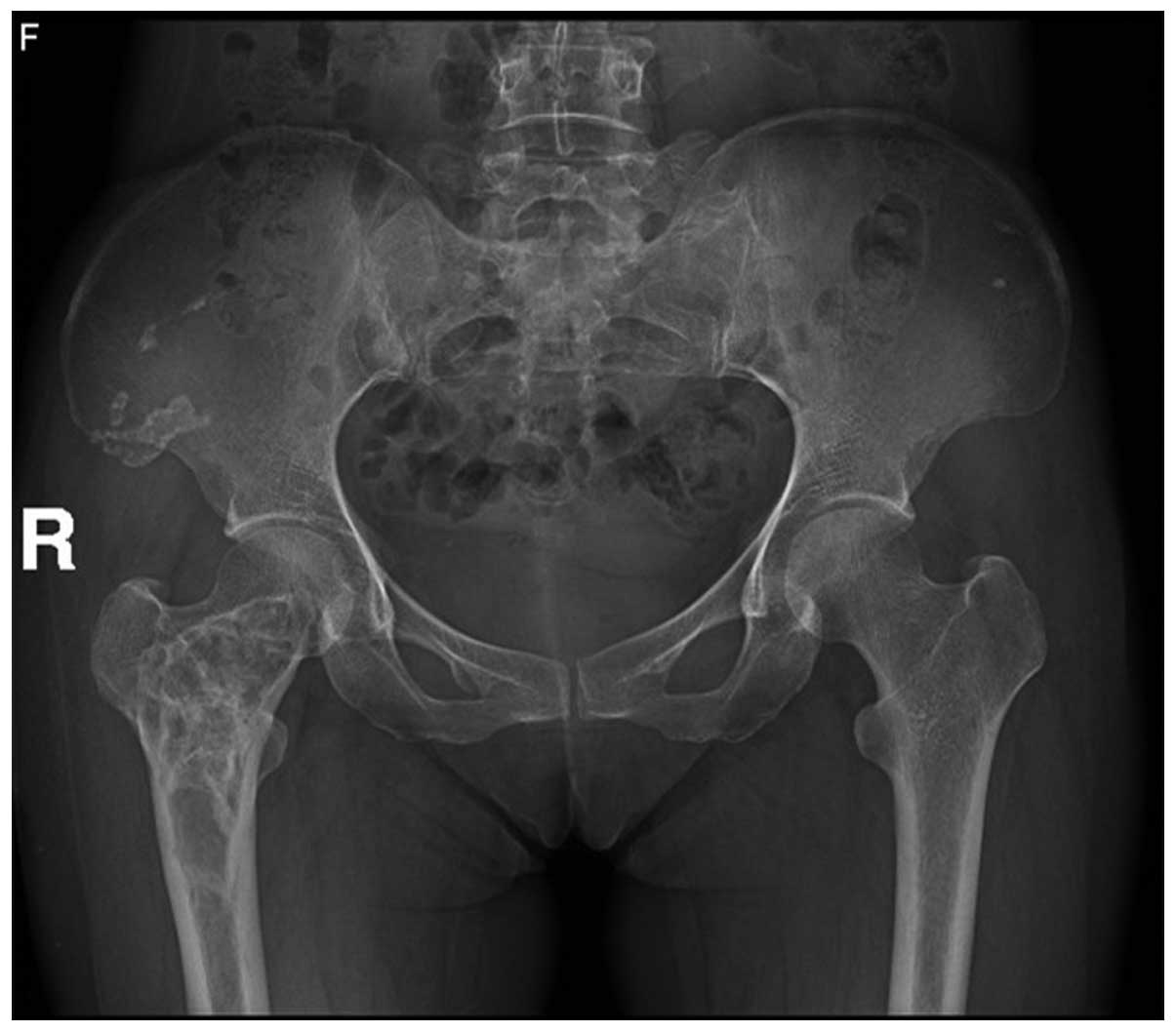
 Radiograph Of Pelvis AP View Depicting Lytic Lesion In Superior Pubic
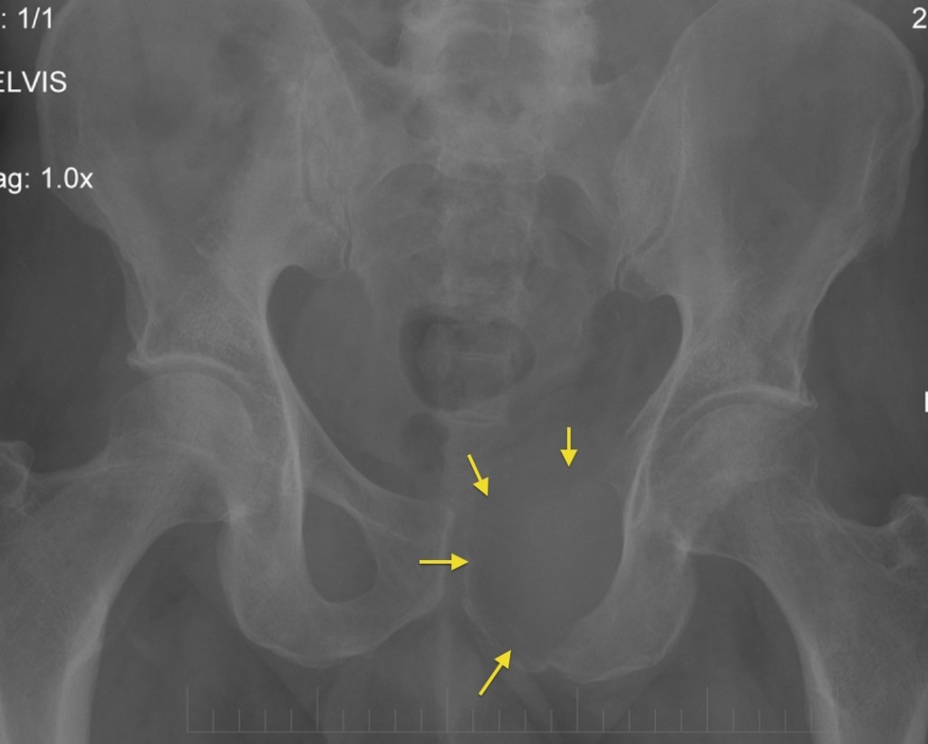 Lytic Bone Metastasis Radiology At St Vincent s University Hospital
 Neuroradiology Spine Lesions Case 4 Overview Choose Your Own
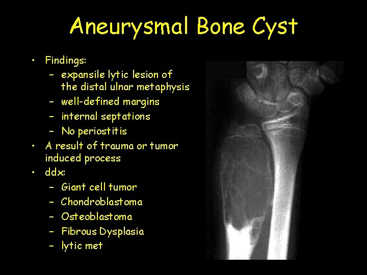 Aneurysmal Bone Cyst Findings Expansile Lytic Lesion Of
 Diagram Of Different Types Of Bone Tumors That Can GrepMed
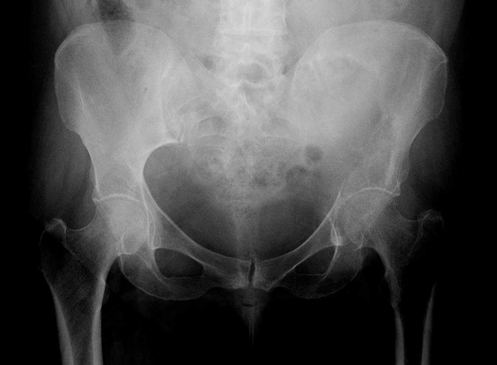 Bone Tumours And Benign Lytic Lesions
 Expansile Nodule Is Located In The Left Lobe Of Thyroid Gland Istmic
 X ray Bilateral Pelvis Showing Lytic Lesion In The Right Iliac Bone
 Stephanie Hart MD On Twitter Patient Presents With A Painless
 What Is TNFRSF11A Gene Osteolysis Familial Expansile NGS Genetic DNA
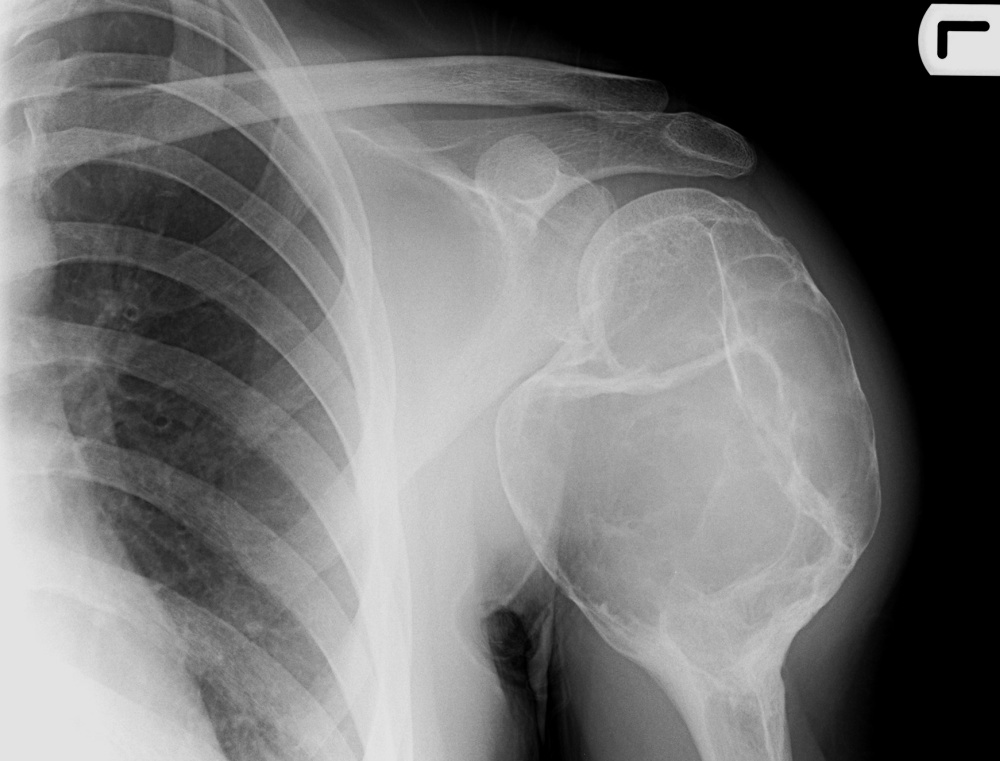 Bone Tumours And Benign Lytic Lesions
 Radiograph Of The Pelvis Showing The Entire Left Ilium With A Large
 Osteosclerotic Bone Tumors Radiology Imaging Medical Anatomy
 Brigham And Women s Pathology On Twitter A Pt Presents W an
 An AP Radiograph Of The Left Scapula Shows A Lucent Expansile And
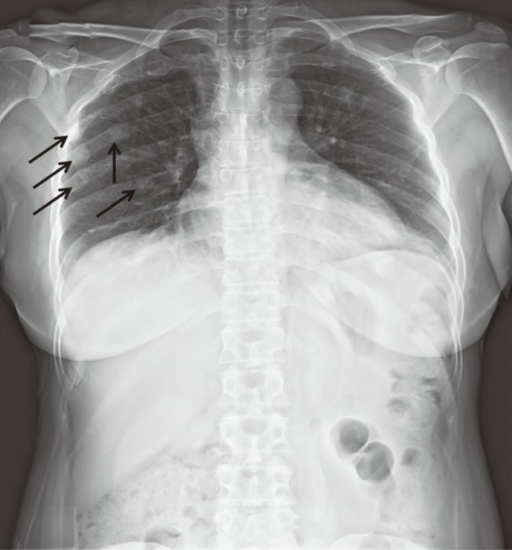 Chest X ray Showed Multiple Sclerotic Lesions Of Right Open i
 Cystic And Cystic Appearing Lesions Of The Mandible Review AJR
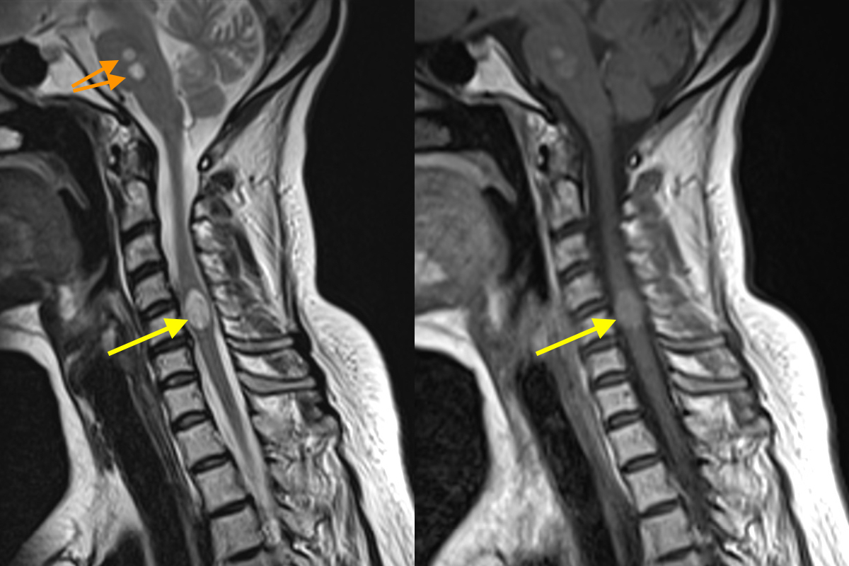
 Lateral Radiograph Of The Cervical Spine A Showing An Expansile
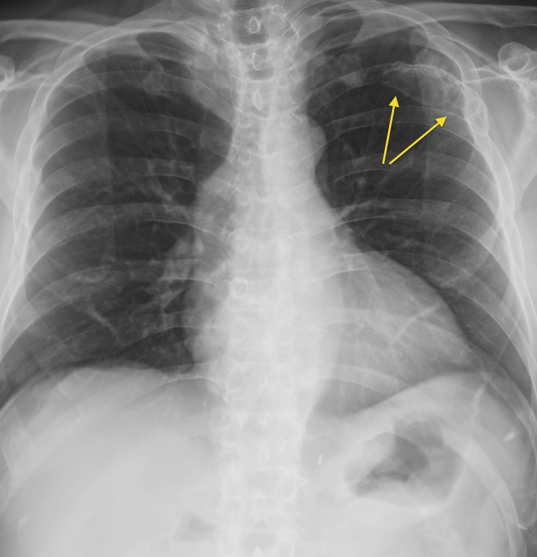 Myeloma In Rib Radiology At St Vincent s University Hospital
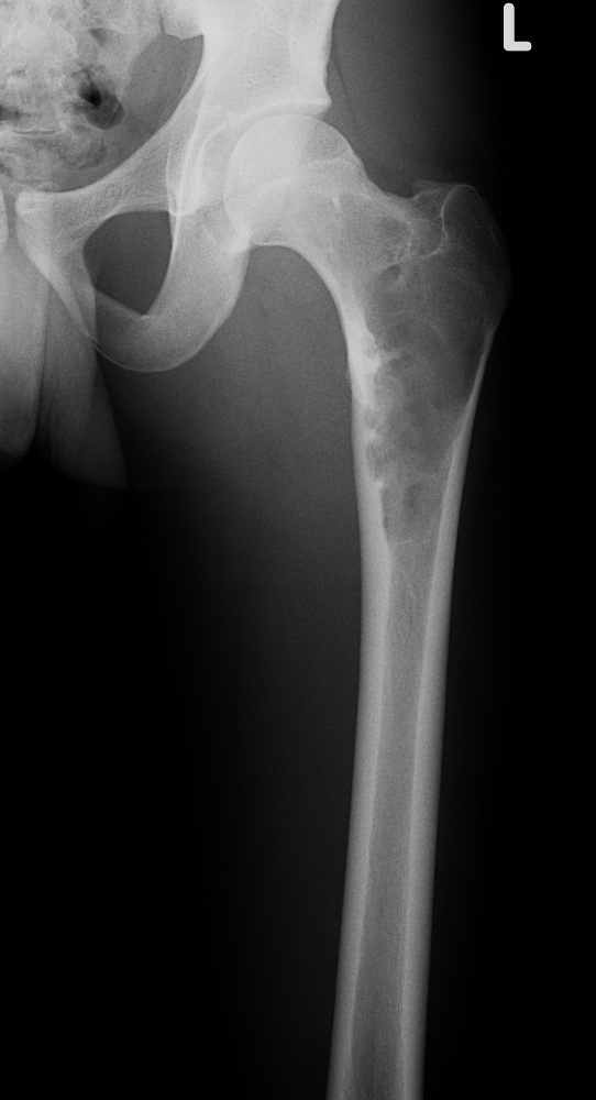 Bone Tumours And Benign Lytic Lesions
 Osteolytic Lesion With Thick Sclerotic Walls And Demyelinating
 JCM Free Full Text Presentation And Prognosis Of Primary Expansile
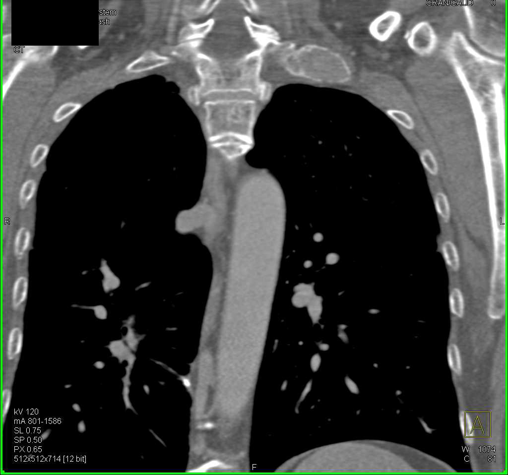 Expansile Rib Lesion Possibly Bone Cyst Post Trauma Musculoskeletal
 Radiodiagnosis Imaging Is Amazing Interesting Cases Giant Cell Tumor
 Dermoscopy Pigmented Lesion On Neck
 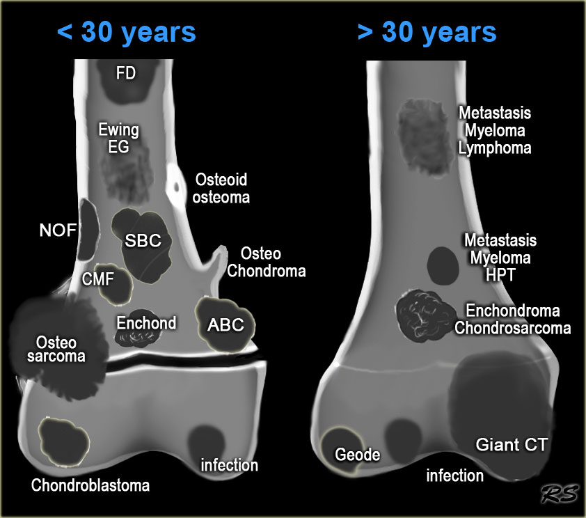 RadiologySpirit Bone Tumors Radiology
 X ray Of The Pelvis Showing Multiple Sclerotic Lesions Over Right Iliac
 A Sclerotic Lesion Is Seen In The Distal Right Femur Of A 37 year OldLung Bone Anatomy
 Clival Epidermoid MRI Sumer s Radiology Blog
 Pin On Radio MSK
 Cureus An Immature Ten year Long standing Case Of Ossifying Fibroma
 Instagram Post By Bhavin Jankharia Jan 19 2017 At 2 43pm UTC
Frequently Asked Questions
Is this Lung Bone Anatomy free to use?
Yes, 100% free. Download and print without creating an account or providing your email address.
What paper size does this template support?
Templates are designed for A4 and US Letter paper. Select 'Fit to page' in your printer dialog for the best fit.
Can I print multiple copies?
Yes. Once you download the image, you can print it as many times as you like for personal or educational use.